Research motivation
Brain image analysis has become one of the main and essential tools for diagnosis, monitoring and prognosis of brain diseases including multiple sclerosis, Alzheimer and stroke. Despite the advances done, the impact on treatment and on care of patients is limited due to the same disease’s complexity and lack of precision of available tools.
ADONIS project
With the ADONIS project (funded by PID2023-146187OB-I00, Spanish PGC 2023 call) we aim to investigate, develop and validate novel and advanced image analysis and deep learning solutions to improve current technology in brain imaging. The project proposes to push beyond the state of the art in different research areas: 1) the development of models for brain age estimation from MRI images that could be used as a biomarker for neurodegenerative diseases; 2) the development of transversal and longitudinal models that could provide more robust change detections; 3) the development of tools for self-supervised and semi-supervised learning, aiming to obtain more robust and generalisable models and to transfer biomarkers and predictive models to different tasks, providing also the capability to perform incremental and continuous learning through the addition of new data; 4) the development of pioneer predictive models fusing the aging biomarker with other relevant clinical data for multiple sclerosis patients; and 5) the development of novel deep learning models for stroke patients to predict patient recanalization (TICI score) from basal CTA images, and to predict hemorrhagic expansion using basal CT images.
MODELLING project
With the MODELLING project (funded by PID2020-114769RB-I00, Spanish RETOS 2020 call) we aim to investigate, develop and validate new computerised tools to quantify brain atrophy and to build predictive models for brain diseases such MS. From the technological point of view, the project pushes the state of the art in various areas, investigating: i) the use of DL techniques to improve current longitudinal MRI brain atrophy biomarkers in the presence of brain lesions; ii) novel DL approaches for image synthesis to generate datasets with controlled/simulated atrophy changes and to deal also with MRI image heterogeneity; iii) development of MS predictive models, investigating both traditional and DL methods to fuse MRI information with other relevant clinical, laboratory and patient data to infer the probability of future events (risk of progression or effectivity of a particular drug); and iv) novel learning to learn approaches to study the applicability of MRI biomarkers and predictive models to different brain diseases such as Stroke or Alzheimer.
EVOLUTION project
With the EVOLUTION project (funded by DPI2017-86696-R, Spanish RETOS 2017 call) we aim to investigate, develop and validate new automated computerised tools to extract MRI biomarkers with the goal of computing MS predictive models that can be applicable in clinical practice. This novel paradigm will provide the basis for improving the prognosis and monitoring of MS, introducing objectivity, and simplifying the everyday practice for radiologists and neurologists. From the technological point of view, the project pushes the state of the art in various areas, investigating: i) the use of deep learning techniques to improve the current MRI brain biomarkers for MS such as brain lesions, brain atrophy, and cortical and subcortical grey matter; ii) novel solutions to deal with the generalisation of these models to different image domains (i.e. different MRI scanners or image resolutions) without requiring large amount of training data; iii) the development of predictive models based on using MRI information such as MS lesion quantification.
BiomarkEM.cat and NICOLE projects
With the BiomarkEM.cat project (funded by "La Fundació La Marató de TV3", neurodegenerative disease call) we aim to develop, validate and implement in clinical practice fully automated and robust tools to measure new lesions and brain volume changes in patients with multiple sclerosis. The coordinated project, with a multidisciplinary team of computer science doctors, radiologists and neurologists with a strong background and experience within the field, will consist in the following essential blocks: data collection, development of tools for obtaining automated MRI-derived estimates and clinical validation.
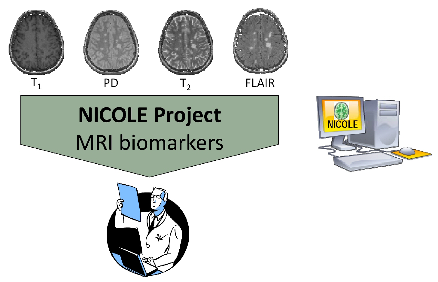
On the other hand, the NICOLE project (TIN2014-55710-R, Spanish RETOS 2014 call) aims to develop new automatic computer tools to extract robust MRI biomarkers . This will enable a new paradigm that will provide the basis for improving the diagnosis and monitoring of MS and stratification of patients by introducing objectivity and simplifying the daily clinical practice.

This NICOLE project aims to advance the state of the art in various areas such as the development of RM biomarkers for MS and the generalization of the tools for use in different MRI machines (Siemens, Philips and General Electric). The data acquired in the project will allow this pioneering work, analyzing the differences in performance with respect to MRI scanners and the development of strategies for obtaining more reliable, robust, efficient and fully automated computerized tools.
Collaboration centers
These two projects are done in collaboration with Hospitals that are reference centers in Catalonia within the Multiple Sclerosis research field. IDIBGI, with Hospital Dr. Josep Trueta and Hospital Santa Caterina from Girona, and with Hospital Vall d'Hebron and Centre d'Esclerosi Multiple (Cemcat) from Barcelona.
Previous projects: SALEM and AVALEM
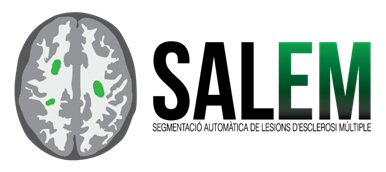
In the SALEM project we developed and validated an automatic system for the detection, segmentation and description of MS lesions based on computer vision techniques. In particular, we aimed to develop a computer aided tool in order to automatically detect and segment the MS lesions in MRI images, and to provide a quantitative description and a qualitative description for each patient.

On the other hand, the AVALEM project aimed to automatically evaluate and quantify the atrophy of the patients and their evolution in time.
These developed tools were tested and evaluated exhaustively in the Hospital centers involved in the projects: Hospital Dr. Josep Trueta, Clinica Girona and Hospital Vall d'Hebron from Barcelona .
Main goal
To introduce improvements in the diagnosis and monitoring of MS and stratification of patients by introducing objectivity and simplifying the daily clinical practice.