Purpose of this website
This webpage provides a summary and installation instructions of software that may be used for segmenting the brain tissue in presence of white matter lesions. The method only requires T1-w images, but its performance is improved if the FLAIR image is also available.
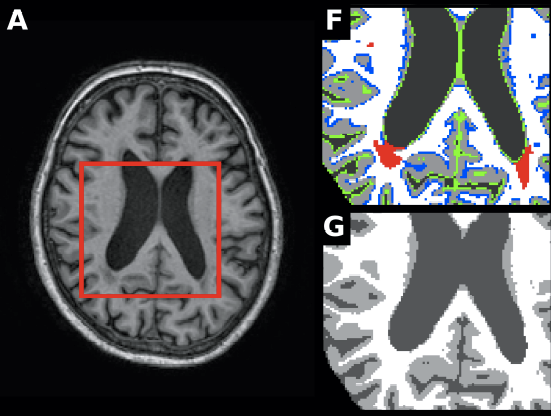
Over the last few years, the increasing interest in brain tissue volume measurements on clinical settings has lead to the development of a wide number of automated tissue segmentation methods. However, white matter lesions are known to reduce the accuracy of automated tissue segmentation methods, which requires manual annotation of the lesions and refilling them before segmentation, which is tedious and time-consuming. MSSEG is a new, fully automated tissue segmentation approach designed to deal with images in the presence of WM lesions. This approach integrates a robust partial volume tissue segmentation with WM outlier rejection and filling, combining intensity and probabilistic and morphological prior maps.a
If you feel interesting our webpage and use some of the resources given, please cite the following paper in your research:
- S.Valverde, A.Oliver, E.Roura, S.González-Villà, D.Pareto, J.C.Vilanova, Ll.Ramió-Torrentà, À.Rovira, and X.Lladó. Automated tissue segmentation of MR brain images in the presence of white matter lesions. Medical Image Analysis, to appear.